Male & Female Dog Reproductive Systems — Organs and Hormones
Dog organs anatomy with the labeled diagram, Male and female dog pelvis anatomy with their organs and labeled diagram. Frequently asked questions on dog abdominal anatomy. Let's see the dog anatomy learner's common inquiries on the dog's abdominal anatomy. All these questions and answers might be helpful for you to learn something more.
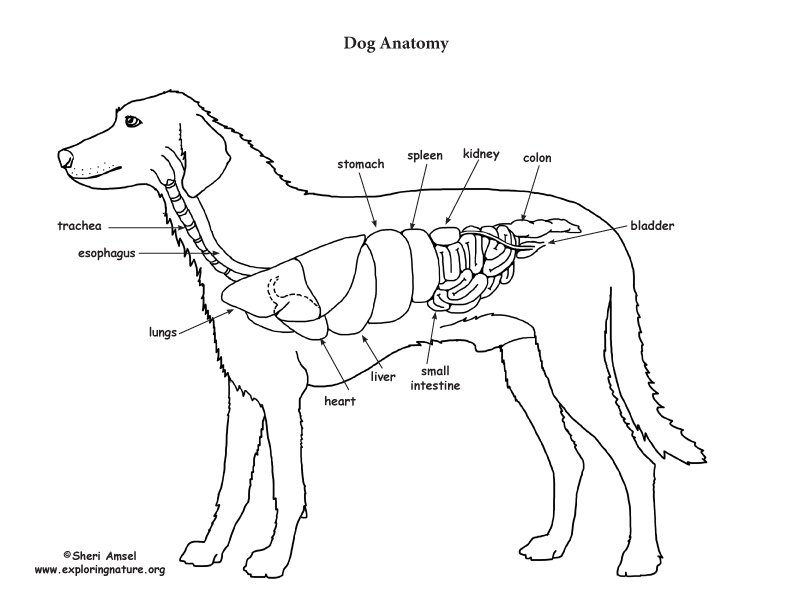
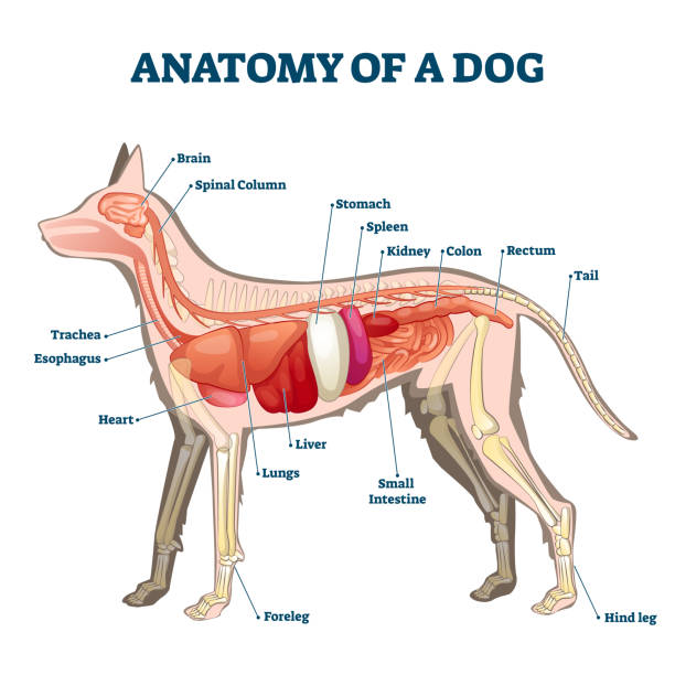
Dog Anatomy (Thoracic and Abdominal Organs)
Dog anatomy comprises the anatomical studies of the visible parts of the body of a domestic dog.Details of structures vary tremendously from breed to breed, more than in any other animal species, wild or domesticated, as dogs are highly variable in height and weight. The smallest known adult dog was a Yorkshire Terrier that stood only 6.3 cm (2.5 in) at the shoulder, 9.5 cm (3.7 in) in length.
Dog Anatomy (Thoracic and Abdominal Organs)
It provides information about a dog's skeletal, reproductive, internal, and external anatomy, along with accompanying labeled diagrams. After mating, dogs experience something called a copulatory tie, wherein they remain in the coital position. The male dog dismounts the female at this time. The dogs can remain in this position from a few.

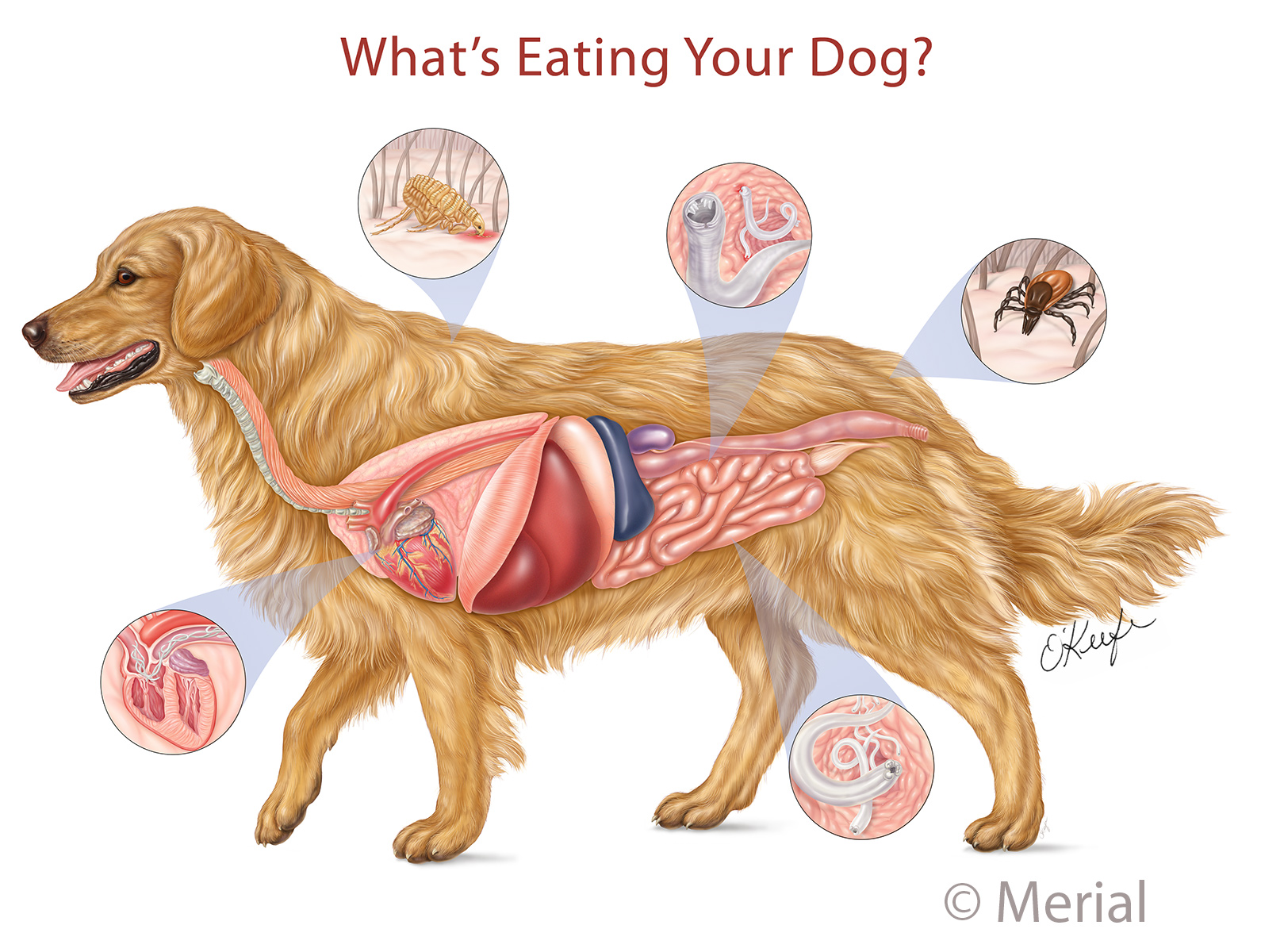
The process of a dog’s digestive system
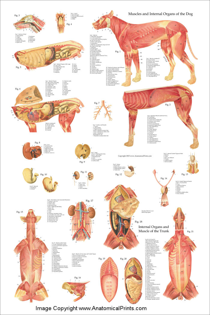
This veterinary anatomy module contains 608 illustrations on the canine myology. Here are presented scientific illustrations of the canine muscles and skeleton from different anatomical standard views (lateral, medial, cranial, caudal, dorsal, ventral / palmar.). Some fascias, tendons, ligaments, joints were labeled.

Canine Internal Anatomy Poster Dog Organs Laminated Chart
Whereas giant breeds can take between 18 months and 2 years for their growth plates to fuse. Speaking of skeletons, a dog has 320 bones in their body (depending on the length of their tail) and around 700 muscles. Muscles attach to bones via tendons. Depending on the breed of dog, they will have different types of muscle fibers.

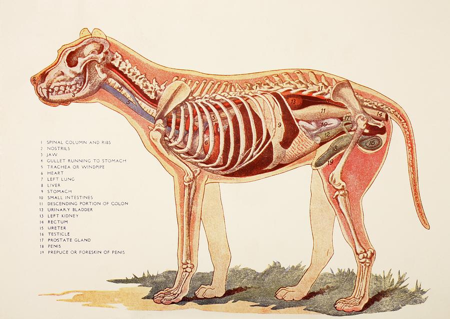
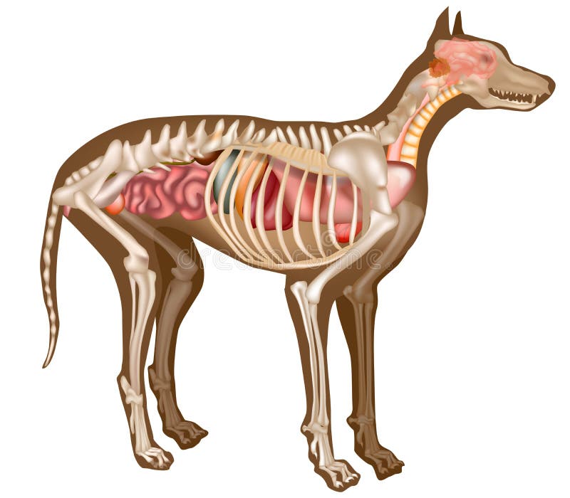
Anatomy of a male dog crosssection, showing the skeleton and internal
The Anatomy of the Canine Digestive System (An Overview) A muscular tube that carries ingesta from the laryngopharynx to the stomach. The esophagus courses through the neck, thorax, and into the abdomen. The esophagus can therefore be referred to as having cervical, thoracic, and abdominal portions. The stomach is a dilation of the alimentary.
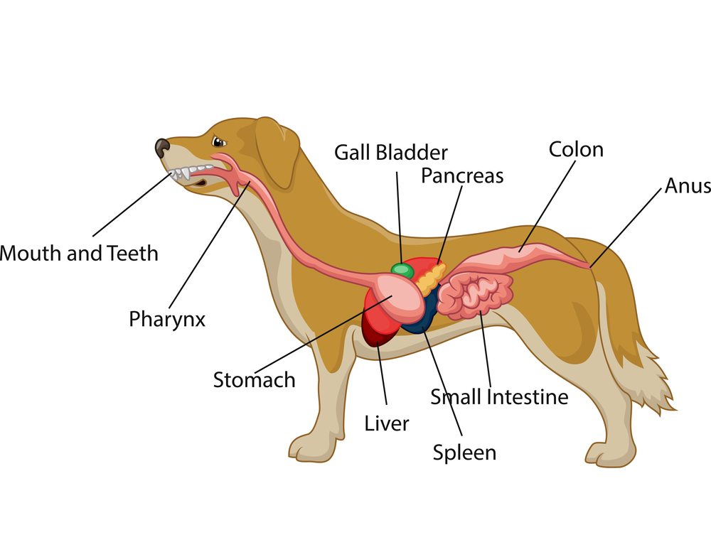
Dog Digestive Process and what the stages are and how it works
The Atlas is not intended to be an exhaustive review of anatomy, pathology, or medicine. For more information, consult the Bibliography, refer to prescribing information on specific drugs, or call Hill's Veterinary Consultation Service at 1-800-548-VETS (8387) or e-mail [email protected]. The Atlas contains illustrations of the most.

Dog Anatomy (Thoracic and Abdominal Organs)
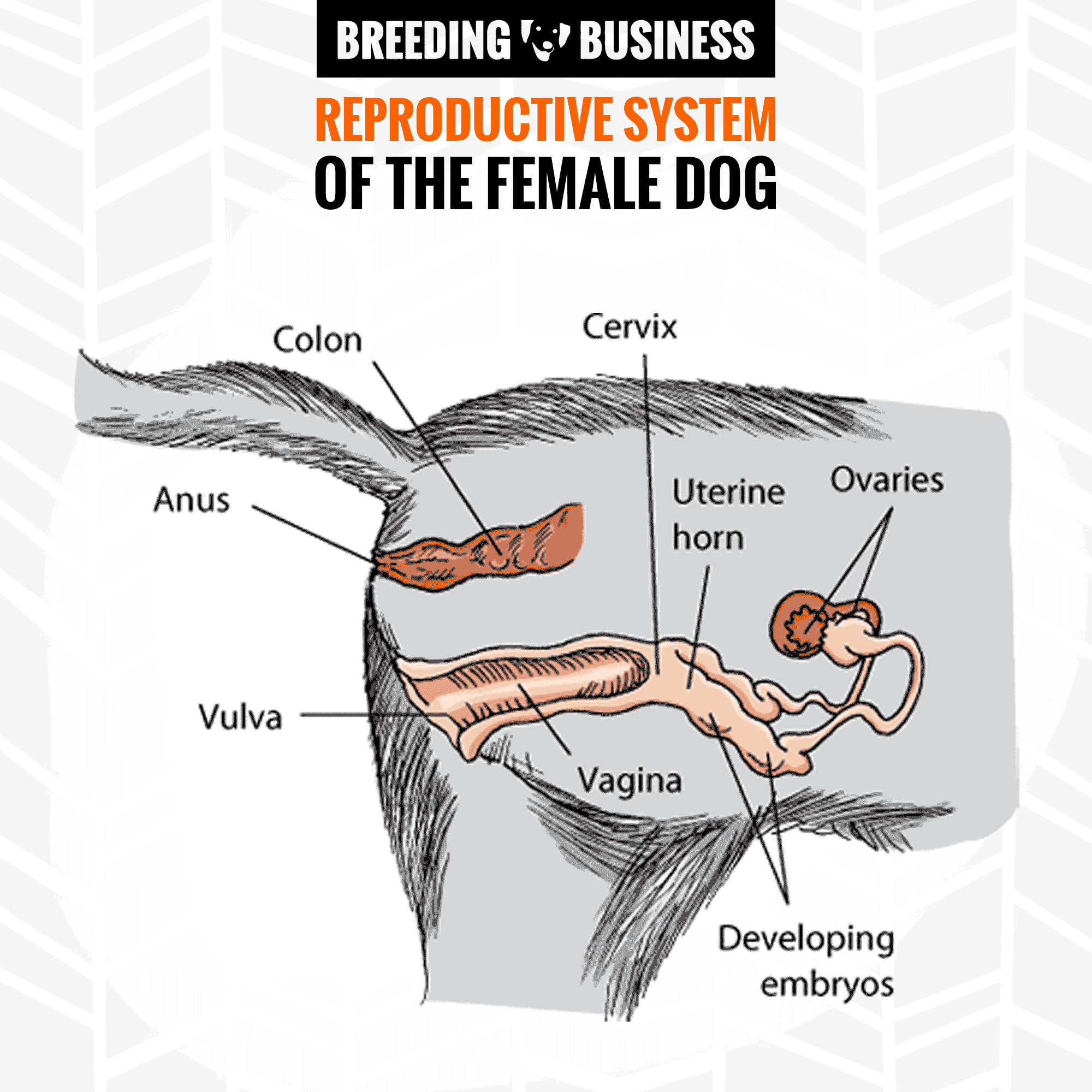
A female dog's reproductive system has similar organs as a human's. The female dog anatomy external organ is the vulva, which opens to the vagina. A pregnant female dog's anatomy includes two ovaries, which produce eggs, the cervix, fallopian tubes, and the uterus. The uterus becomes the womb for her puppies during their gestation period.
Dog Internal Anatomy Anatomical Charts & Posters
Anatomy atlas of the canine general anatomy: fully labeled illustrations and diagrams of the dog (skeleton, bones, muscles, joints, viscera, respiratory system, cardiovascular system). Positional and directional terms, general terminology and anatomical orientation are also illustrated.
Internal Organs Of A Male Dog. From Photograph by Ken Welsh Pixels
When you research information you must cite the reference. Citing for websites is different from citing from books, magazines and periodicals.
Coughing in Dogs May Signal Heart Disease HubPages
Xiphoid region (Cranial abdominal region) Zygomatic bone. Zygomatic gland. Zygomatic region. Radiographic anatomy: labeled images in the transverse plane of a healthy dog's whole body, using tomodensitometry. Introduction to the anatomy of the skull, thorax, abdomen, pelvic cavity, muscles and blood vessels: main anatomical structures identified.
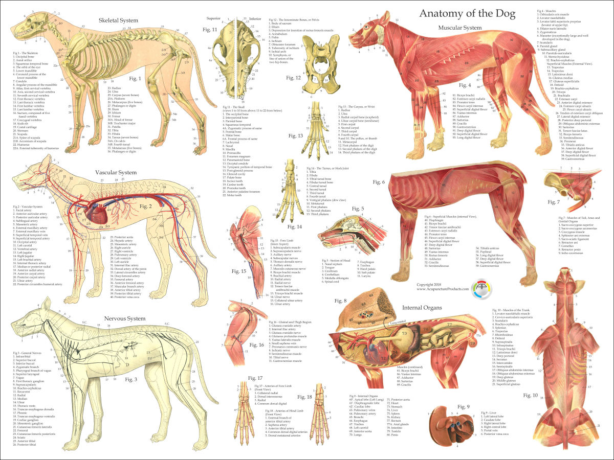
Dog Anatomy Poster
ISSN 2534-5087. This vet-Anatomy module presents an anatomy atlas of the abdomen and pelvis of the dog in CT. CT images are from a healthy 6-year-old castrated male dog. This module displays cross-sectional labeled anatomy images of the canine abdominal cavity and the pelvis on a Computed Tomography (CT) and 3D images of the abdomen of the dog.
Dog Internal Anatomy Poster 24 x 36
The Anatomage Dog is the first highly detailed dog anatomy atlas that comprehensively features internal organs, including vascular systems and muscular-skeletal structures. Originating from real dog data, the Anatomage Dog exhibits the highest level of anatomical accuracy. All of its volumetric 3D and individual structures are segmented, users.
Dog Anatomy Stock Photos, Pictures & RoyaltyFree Images iStock
Dog skeleton. As with any vertebrate animal, the skeleton of a dog has the function of supporting the body for movement and protecting its internal organs. We can divide the canine skeleton into three main sections: Axial skeleton: skull, spine, ribs and sternum bones. Appendicular skeleton: bones of the extremities.

Anatomy Of Back Organs / Anatomy Male Organs in Loop Stock Footage
This canine internal anatomy poster illustrates the internal anatomy of a dog in beautifully rendered detail. This dog organ anatomy poster has been designed exclusively for AnatomyStuff and is medically accurate, making it the perfect choice for display in a veterinary classroom or practice, or in a vet clinic for owner education.. This canine organs chart offers the following features:
Canine Internal Anatomy Chart. Anatomy of Dog with Inside Organ
Dog Anatomy Flash Cards by Bryan Edwards Publishing The Dog Anatomy is a comprehensive reference tool covering the skeletal system, muscular system, joints & ligaments, and the 10 major organ systems of the dog. This set consists of 47 flash cards.
